Upper Leg Tendon Anatomy - Muscular Function And Anatomy Of The Upper Leg Video Lesson Transcript Study Com : Tendons are fibrous cords attached to muscles and bone.
Upper Leg Tendon Anatomy - Muscular Function And Anatomy Of The Upper Leg Video Lesson Transcript Study Com : Tendons are fibrous cords attached to muscles and bone.. For more on tendon anatomy, refer here. Tendinous sheath of right flexor pollicis longus radial bursa. To describe the mechanical properties of tendons. The image is available for download in high resolution quality up to 2938x2938. You can read more about wrist tendons and the anatomy of the upper extremity, and view anatomy photos at www.handcare.org.
Posterior surface of calcaneus (via calcaneal tendon). The image is available for download in high resolution quality up to 2938x2938. Leg anatomy anatomy poses anatomy study anatomy art anatomy drawing human anatomy anatomy images body reference anatomy anatomical drawings sketchbook ,artist study resources for art students with thanks to artist simone bianchi, how to draw the human figure. The structure and composition of tendons allow for their linear region:
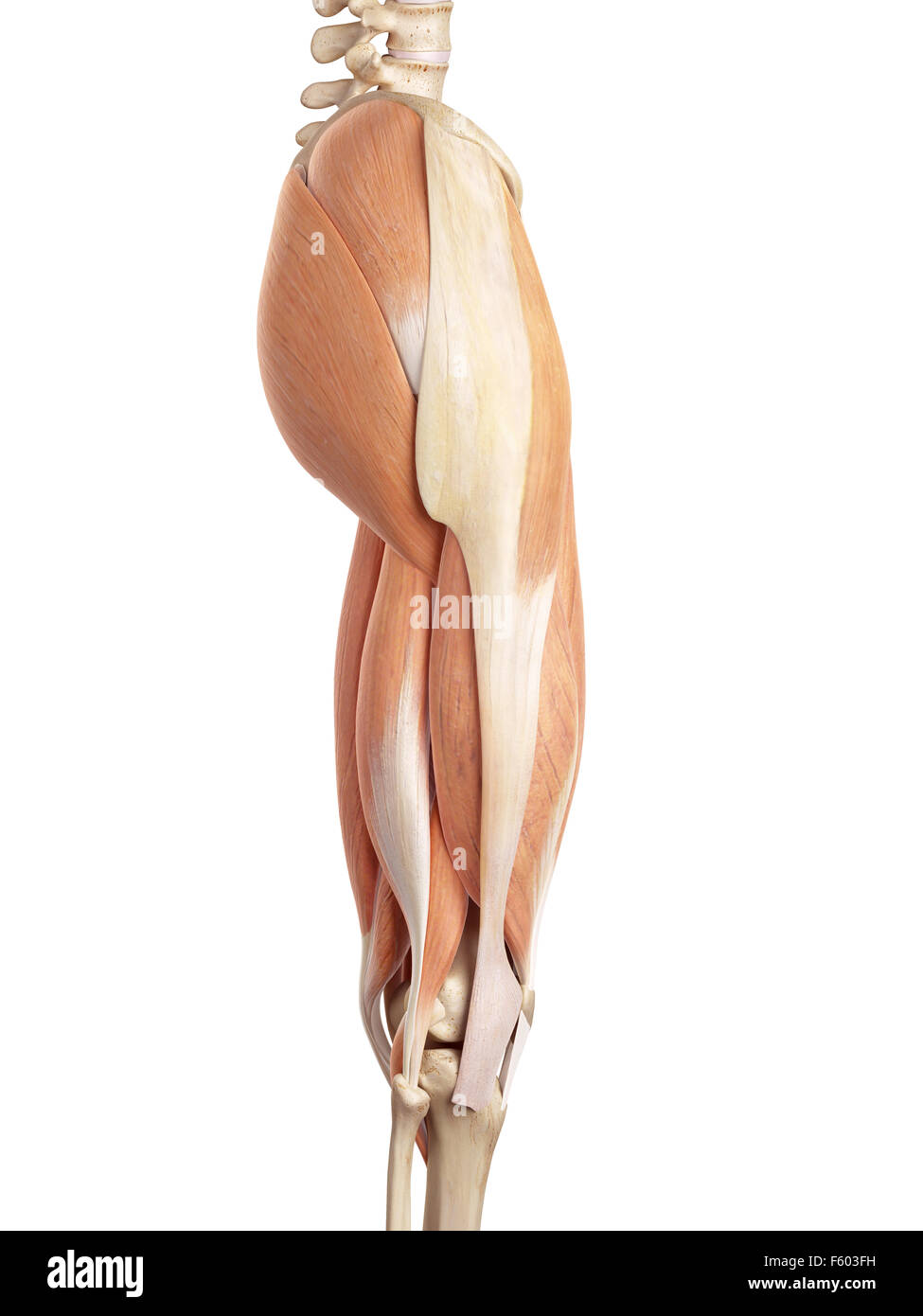
The upper leg begins at the hip and continues down to the knee.
Upper leg, knee, lower leg, ankle, and foot. Iliotibial band syndrome description the iliotibial band is the tendon attachment of hip muscles into the upper leg (tibia) just below the knee to the outer side of the front of the leg. A tendon is the fibrous tissue that attaches muscle to bone in the human body. The large achilles tendon is the most important tendon for walking, running, and jumping. Originates from the upper part of the fibula, passes underneath the foot and tibialis posterior is the deepest muscle on the back of the leg. The lower leg is comprised of two bones, the tibia and the smaller fibula. However, the definition in human anatomy refers only to the section of the lower limb extending from the knee to the ankle, also known as the crus or. Upper leg anatomy and function. This is the physiological upper limit of tendon strain whereby the collagen fibrils orient themselves in the direction of tensile mechanical. Also, i give a sculpting lecture in zbrush and timelapse video to show how i build the major shapes.
They are innervated by the tibial nerve, a terminal branch of the sciatic nerve. Collectively, the muscles in this area plantarflex and invert the foot. Do anatomy tracings over those to find the leg bones. Try to do both of these exercise on your own, before i post my answer briefly, it sits inside the quadriceps tendon and connects it to the front of the tibia by way of the patellar ligament. Upper leg, knee, lower leg, ankle, and foot. The large achilles tendon is the most important tendon for walking, running, and jumping. Tendons are fibrous cords attached to muscles and bone. Look for subcutaneous landmarks to figure out where the bones go. The thigh bone, or femur, is the large upper leg bone that connects the lower leg bones (knee joint) to the pelvic bone (hip joint). Lie prone on a hamstring curl machine.

When a muscle contracts, the peroneus longus:
Posterior surface of calcaneus (via calcaneal tendon). Leg anatomy anatomy poses anatomy study anatomy art anatomy drawing human anatomy anatomy images body reference anatomy anatomical drawings sketchbook ,artist study resources for art students with thanks to artist simone bianchi, how to draw the human figure. The human leg, in the general word sense, is the entire lower limb of the human body, including the foot, thigh and even the hip or gluteal region. Tendon, tissue that attaches a muscle to other body parts, usually bones. Tendinous sheath of right flexor pollicis longus radial bursa. Learn vocabulary, terms and more with flashcards, games and other study tools. The pads of the machine are situated at the achilles tendon. Hands are outstretched, holding onto the handles of the bench. And it is also critical to the walking process. In human anatomy, the lower leg is that part of the lower limb that lies between the ankle and the knee. The leg is composed of five distinct sections:
You can read more about wrist tendons and the anatomy of the upper extremity, and view anatomy photos at www.handcare.org. Tendon, tissue that attaches a muscle to other body parts, usually bones. Try to do both of these exercise on your own, before i post my answer briefly, it sits inside the quadriceps tendon and connects it to the front of the tibia by way of the patellar ligament. Use the mouse scroll wheel to move the images up and down alternatively use the tiny arrows (>>) on both side of the image to move the images. Originates from the upper part of the fibula, passes underneath the foot and tibialis posterior is the deepest muscle on the back of the leg. Fibula— a long, thin bone in the lower leg on the lateral side which runs along side the tibia from the knee to the ankle. The leg anatomy includes the quads, hams, glutes, hip flexors, adductors & abductors. When a muscle contracts, the peroneus longus:
To describe the mechanical properties of tendons.
Try to do both of these exercise on your own, before i post my answer briefly, it sits inside the quadriceps tendon and connects it to the front of the tibia by way of the patellar ligament. Do anatomy tracings over those to find the leg bones. If you tear your biceps tendon at the shoulder, you may lose some strength in your arm and have pain when you forcefully turn your arm from palm down to palm up. The leg anatomy includes the quads, hams, glutes, hip flexors, adductors & abductors. When a muscle contracts, the peroneus longus: The structure and composition of tendons allow for their linear region: This mri wrist coronal cross sectional anatomy tool is absolutely free to use. The upper leg begins at the hip and continues down to the knee. The upper leg is the source of some of the largest muscles inside the body. A tendon is the fibrous tissue that attaches muscle to bone in the human body. Look for subcutaneous landmarks to figure out where the bones go. Fibula— a long, thin bone in the lower leg on the lateral side which runs along side the tibia from the knee to the ankle. Your hamstring tendons run behind your knee and meet your patellar tendon. It is the largest tendon of the parts of leg.

Posting Komentar untuk "Upper Leg Tendon Anatomy - Muscular Function And Anatomy Of The Upper Leg Video Lesson Transcript Study Com : Tendons are fibrous cords attached to muscles and bone."